The impact of robotics on neurosurgery
11th October 2016
Stuart Campbell, Clinical Sales Development Manager of the Neurological Products Division at Renishaw, discusses key trends on the use of robotics in neurosurgery.
The curious case of Phineas Gage is one of the earliest and best known cases of serious brain injury. On September 13th, 1848, Gage was working as a railway foreman in Vermont when an explosion caused a three foot long iron rod to be propelled straight through his skull. At the time, doctors thought it impossible to survive such an injury and his remarkable survival and reported personality changes affected the study of neuroscience forever. In recent years, a new technology has changed the face of neuroscience - robotics, which offers high precision access to a complex and sensitive region.
Industrial environments are rife with automation and robotic systems. The upwards trend is only increasing, with the International Federation of Robotics predicting that by 2018, 1.3 million industrial robots will be entering service in factories across the globe. Automated or robotic systems can increase the speed, reliability and accuracy of industrial processes, but the benefits of robotics are not limited to industrial applications.
Applications in the operating theatre
The first application of a robotic system in surgery happened in 1985, 24 years after the introduction of UNIMATE, the first industrial robot. In this first robotic surgery, surgeons performed a neurosurgical biopsy using a PUMA 560 robotic arm. The robotic system allowed for greater precision in minimally invasive surgery compared to more traditional methods.
Despite the first application of a robot assisted procedure being in neurosurgery, robotic systems are not as widely used in this field compared to other areas of medicine such as urology, cardiology and gastroenterology. This is partly because of the anatomical challenges in such a complex and spatially limited organ but also because of the fact that the brain includes extremely sensitive tissue.
Brain tissue may be difficult to access and manipulate, but it is also incredibly important. As a result, technological improvements to traditional methods have always been a focus to improve the precision of surgery and release the potential of the technology in this important area of the body. Advances in engineering and imaging techniques have sparked further interest in computer-assisted and robotic neurosurgery.
The need for precision during brain surgery has led to an increase in computer-assisted surgeries (CAS). This technique involves using imaging technologies such as magnetic resonance imaging (MRI), computerised tomography (CT) or positron emission tomography (PET) to generate an image of the patient's brain. The surgeon will use this information to plan the route of surgery.
Computer-assisted surgery can accurately guide surgeons to their surgical targets, therefore improving patient outcomes by limiting damage to adjacent tissue. CAS has been a key factor leading to robotic-assisted surgery. It enables the surgeon to use software to control and move surgical instruments mounted on a robot and perform surgery through small incisions.
Surgical robots
One definition of a surgical robot is ‘any reprogrammable powered manipulator with artificial sensing'. The most important factors to consider when classifying a surgical robot are in its surgical applications, its level of interaction with the surgeon and the role of the robot in the surgery. Robots range from being fully dependent, where the surgeon has full control of the system for the duration of the procedure, to autonomous, where the robot will reproduce pre-programmed motions or instructions during the surgery without the control of the surgeon.
Currently, the most common systems in robotic surgery are dependent systems, where the surgeon retains full control of the surgical instruments. This type of surgery is also known as telesurgery and a popular example of a telesurgery robot is the da Vinci® Surgical System which enables surgeons to perform minimally invasive surgery. It provides a 3D HD view, ergonomic design and wristed instruments that can bend and rotate more than the human hand.
Surgeons have used this system in over three million surgeries since it received FDA approval in 2000. Autonomous systems are less common, but surgeons do currently use this equipment in stereotactic neurosurgery, a sector where robotic systems are growing in popularity.
Stereotactic surgery
Stereotactic neurosurgery is a technique used by neurosurgeons to locate surgical targets within the brain. It uses 3D imaging data and either an external frame or imaging markers attached to the scalp as reference points. This technique enables surgeons to reach targets that are deep in the brain in a minimally invasive way. Surgeons would most commonly use this technique in procedures including deep brain stimulation (DBS), stereoelectroencephalography (SEEG), biopsy and endoscopy, or to deliver devices or instruments to a small target in the brain.
Traditionally, in frame-based stereotactic surgery, the surgeon would attach a frame to the patient's head and use an imaging technique to identify the best routes to the target area. The frame provides a fixed support to accurately position surgical instruments according to 3D coordinates.
Surgeons must use the imaging information to identify the most suitable angle to enter the brain in order to minimise the risk of damaging vital tissue. In a brain biopsy, the target coordinates help to position and pass a probe through a small hole in the skull. In another example, in the main type of procedure to treat the symptoms of Parkinson's, electrodes are placed deep in the brain to deliver high frequency stimulation.
Robotics in stereotactic neurosurgery
The first commercially available neurorobotic device for stereotactic neurosurgical procedures was the neuromate® stereotactic robot. This device can decrease procedure time and increase safety in stereotactic neurosurgery in frame and frameless procedures. The robot has five degrees of freedom, can be mounted with surgical instruments and can be used in various procedures.
Surgeons have used the neuromate in thousands of electrode implantation procedures for DBS, SEEG, neuroendoscopy and biopsies. It is now used in many hospitals around the world with several installed in the UK.
Thanks to developments in medical IT, there is now easy to use procedure planning software such as Renishaw'sneuroinspire™ for stereotactic procedures. Integration of the software with the robot provides the surgeon with a system where the programmed robot can be positioned to enable the surgeon to place instruments and/or devices into the correct location. This is effective in reducing human errors and operating time.
Training surgeons in robotics
One of the challenges to the widespread acceptance of robotics in the neurosurgical operating theatre, once they have been proven effective and safe, is the ability to train neurosurgeons to use the innovative technologies.
Simulation techniques are continually improving and the more lifelike the simulation the better, as simulation is a good alternative to cadavers. A benefit of a simulation is that it can be specific to an individual patient if generated using preoperative imaging, so a surgeon can prepare and practice an exact patient-specific procedure using the technology as a dry run. Improvements in virtual reality techniques will prove useful in training surgeons to use technology of the future.
It is extremely important that surgeons have a familiar and comfortable environment in which to practice using Renishaw's technology. This is one of the reasons why Renishaw has set up a Healthcare Centre of Excellence at its Miskin site, near Cardiff in Wales. Within this centre, there is a mock operating theatre suite that mimics a real-life hospital setting, but without the complication of a sterile environment. In the state-of-the-art suite, surgeons can be trained to perform highly complex stereotactic procedures using the Renishaw range of neurological products.
Another technology making its mark on surgical training is live streaming. The first live streamed surgery was broadcast in April 2016 to medical students as well as other interested parties. Live streaming has broken geographical barriers so that experienced surgeons can demonstrate surgical techniques and procedures in real time. The Renishaw Healthcare Centre of Excellence includes this technology in its mock operating theatre.
In the neurosurgical field advances will continue to improve speed, tactile ability and human-robot interfaces. Completely autonomous surgery is still a long way off, but robotics is already changing the face of neurosurgery forever, although in a slower, more progressive way than the explosion that affected poor Phineas Gage.
Downloads
-
News release: The impact of robotics in neurosurgery
Stuart Campbell, Clinical Sales Development Manager of the Neurological Products Division at Renishaw, discusses key trends on the use of robotics in neurosurgery.
[113kB] -
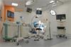 Operating Theatre at the Healthcare Centre of Excellence
The operating room in Miskin is a demo facility used to display Renishaw's range of products for stereotactic neurosurgery
[7.0MB]
