inVia™ Raman microscope aids discovery of rare mineral in alpine plants
August 2018
A team of scientists at the Sainsbury Laboratory Cambridge University (SLCU) have discovered a rare mineral in alpine plants. Vaterite, a form (polymorph) of calcium carbonate, holds enticing potential as a new material for industrial and medical applications. This is the first time that vaterite has been found in plants.
The discovery of vaterite was made through a collaboration between SLCU and Cambridge University Botanic Garden as part of an ongoing research project that is probing the inner workings of plants in the Garden using new microscopy techniques, including Raman spectroscopy.
Dr Raymond Wightman, the Microscopy Core Facility Manager at SLCU's microscopy facility, has a Renishaw inVia™ Raman microscope which is used for studying aspects of plant development and plant cell biology. He said: “This alpine work is just one small part of a number of projects that look at the inner workings of plants. We use the inVia microscope to identify and map molecules.”The microscopy facility consists of general light, confocal and electron microscopes. Dr Wightman said: “Our cryo scanning electron microscope (cryoSEM) allows us to view, in great detail, cells and plant tissues in their ‘native' fully hydrated state by freezing samples quickly and maintaining cold under a vacuum for electron microscopy. The inVia is a unique capability that allows us to probe the biochemistry of plant tissues, cells and even subcellular compartments. It is good for live imaging; we can put a fresh sample under the microscope and get a spectrum within seconds. When the inVia was installed we were delving into the unknown as the lab has never used any other Raman systems. The inVia gives us new information compared to the dynamics and ultrastructural characterisation offered by the facility's high-end confocal and electron microscopes.”
The Renishaw inVia is integrated with a confocal laser scanning microscope (CLSM), allowing correlated Raman and fluorescence measurements. Dr Wightman is also investigating correlative FLIM-confocal-Raman microscopy for observing protein-protein interactions, 3D cellular organisation and distribution of molecular species.
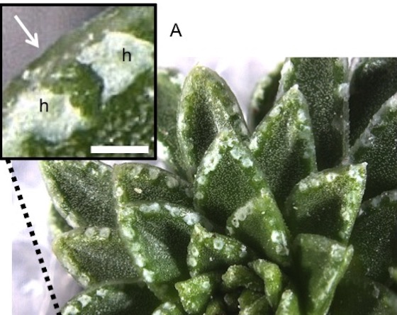
The Garden has a national collection of European Saxifraga plant species. Vaterite was found to be a dominant component of the protective silvery-white crust that forms on the leaves of a number of alpine plants. Dr Wightman said: “In this case, the inVia microscope not only identified signatures corresponding to calcium carbonate as forming the crust, but was also able to differentiate between the calcite and vaterite forms when it was present as a mixture while still attached to the leaf surface.”
This discovery is all the more exciting because naturally occurring vaterite is rarely found on Earth. This is the first time that it has been found in such a large quantity and the first time it has been found to be associated with plants.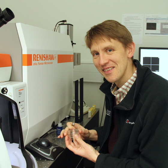
The collaborative work of the SLCU and Cambridge University Botanic Garden team is revealing fascinating insights into leaf anatomy and biochemistry as well as demonstrating the potential for Saxifrages to supply a new range of biomaterials.
More information about the collaboration's discovery of vaterite can be found at www.slcu.cam.ac.uk/news/vaterite-found-in-plants-for-first-time
Images of plants, courtesy of Paul Aston. Image of Dr Wightman, courtesy of SLCU.Downloads
For further images, videos, company biographies or information on Renishaw and its products, visit our Media Hub.