현재 사용 중인 언어로는 이 페이지를 사용할 수 없습니다. Google Translate을 사용하여
자동 번역된 페이지
를 볼 수 있습니다. Renishaw에게는 이 서비스를 제공할 책임이 없으며 번역 결과를 저희가 확인하지도 않았습니다.
추가로 도움이 필요하시면
저희에게 연락해 주십시오.
Geologists use Renishaw's Raman-in-SEM solution to probe the nanoworld
March 2017
Researchers at BRGM (the French Geological Survey in Orleans, France) study the physical, chemical, and structural properties of minerals. BRGM is a public agency which produces and disseminates geological information for government departments, foreign countries, industry and academic research in France and across the world. They use a co-located SEM-Raman system from Renishaw to provide comprehensive in situ sample characterisation.
Dr Guillaume Wille works in the Mineral Physicochemical and Textural Characterization Unit. In describing the analysis methods used at BRGM, Dr Wille said, “Numerous techniques are used, like infrared spectroscopy, X-ray diffraction, electron microscopy and microanalyses (SEM-EDS, TEM-EDS and EPMA), as well as Raman spectroscopy.”
BRGM facilities include a SEM-Raman instrument consisting of a Renishaw inVia™ confocal Raman microscope coupled to a TESCAN Mira3 scanning electron microscope via Renishaw's Structural and Chemical Analyser (SCA) interface. Dr Wille commented, “With this innovative equipment, we have revealed structures with Raman spectroscopy at a nano/micrometric scale thanks to the many imaging and mapping modes of SEM. With the classical optical coupling mode, the optical contrast and resolution are not sufficient. In other words, Raman spectroscopy takes advantage of the nanoresolution of SEM, a powerful advantage when working with nanostructured materials.”
Dr Wille continued, “Examples of work where we use SEM-Raman include: probing submicrometric phases (solid inclusions) and particles inside a matrix; Raman analyses of heterogeneous particles that can only be discriminated by the SEM's contrast or mapping modes as nanofibers; airborne particles discrimination (SE, BSE, CL); and characterisation of powders, where the SCA interface allows the analysis of the same micrometer-sized particle, which would not be possible if the sample had to be transferred from the SEM to the Raman microscope.”
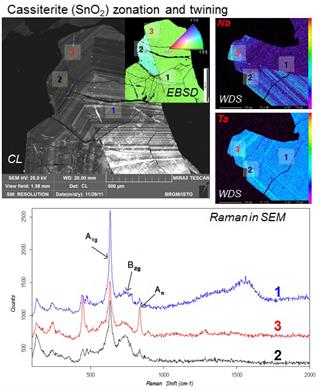
The work carried out at BRGM covers a wide range of applications, with recent examples including environmental asbestos investigation (natural nanofibers), identification of contaminants in soils and aerosol particles, and studying cassiterite (tin oxide) by Raman spectroscopy and cathodoluminescence.
The work of Dr Wille and his colleagues is well reported in several publications. Notable publications include ‘Coupled SEM-microRaman system: A powerful tool to characterize a micrometric aluminum-phosphate-sulfate'1, which reports how the SEM-SCA was used to analyse micrometric aluminum-phosphate–sulfate (APS) occurring in complex geological systems, such as Martian samples. Tiny grains with complex variable composition were simultaneously analyzed in SEM (BSE imaging), EDS and Raman-in-SEM spectroscopy.
The second paper, ‘Raman-in-SEM, a multimodal and multiscale analytical tool: Performance for materials and expertise'2, details studies of the metrological aspects of the use of Raman in the SEM and the use of multimodal analyses, coupling imaging, microanalysis EDS and EBSD, and Raman.
Renishaw's SCA interface uses retractable optics to position the Raman analysis point in the centre of the SEM. This allows both cathodoluminescence and Raman information to be obtained. The system simultaneously acquires SEM and Raman data from the same point on the sample without having to move position or transfer the sample to a different instrument. Both the SEM and inVia can be operated as stand-alone systems, at the same time, without compromising the performance of either.
Please visit www.renishaw.com/SEMRaman for further details on how Renishaw's inVia and SCA interface is being used for co-located analyses.
References
1. N. Maubec et al: Coupled SEM-microRaman system: A powerful tool to characterize a micrometric aluminum-phosphate-sulfate, Journal of Molecular Structure, 1048, 33-40 (2013). DOI: 10.1016/j.molstruc.2013.05.019
2. G. Wille et al: Raman-in-SEM, a multimodal and multiscale analytical tool: Performance for materials and expertise, Micron 67, 50-64 (2014). DOI: 10.1016/j.micron.2014.06.008
Downloads
For further images, videos, company biographies or information on Renishaw and its products, visit our Media Hub.


