라만 분광기로 생물학적 조직 분석
비침습적, 비표지 기법인 라만 분광기는 조직 분석에 적합한 분석 도구입니다.
생체 분자, 마커, 착색 또는 다이를 타겟으로 할 필요 없이 전체 화학 정보 스펙트럼(핵산, 단백질, 지질 등의 물질로부터)을 추출할 수 있습니다. 웨스턴 블롯, GC/MS, MALDI-TOF 등의 많은 다른 분석 기법과 달리 라만 분석에서는 샘플을 균질화할 필요가 없습니다.
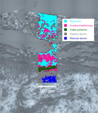
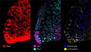
신속하고 정확하게 조직 계층 식별
전암 조직과 암 조직, 건강한 조직을 구별, 식별 및 구분
- 항체와 결합할 필요 없음: 프로토콜을 최적화할 때 금전과 시간을 절약할 수 있습니다.
- 총 분자 조성을 기준으로 조직에서 해부학적 계층을 확실하게 구별하고 객관적으로 식별 종양 변연 설명
- 주관적 비색법과 형태 기준 분석 회피
- 형태학적 변화로 표출되지 않는 조직의 화학적 변화(예: DNA/RNA, 글리코겐, 지질, 단백질, 지질 상 및 DNA 무결성 수준) 식별
생물학적 계통 이해
조직의 완벽한 화학적 분석과 변화의 기본 메커니즘 이해
연구:
- 유기체 성장
- 질병의 발병
- 마약 또는 각성제(예: 화학요법제, 독소, 항염증제)에 보이는 조직의 반응
한 가지 절차로 다음 사항을 연구하십시오.
- 단백질, 지질, 핵산, 탄수화물의 함량 및 구조 변화
- 무기질 침착물의 존재(예: 유방 조직의 석회화, 죽상 동맥 경화증)
- 헴 단백질의 산화/환원 상태(예: 뉴로글로빈 및 미오글로빈)
이미지 조직
Renishaw의 StreamLine™ 기술은 조직 이미지 생성에 특히 적합합니다. 선형 초점 형상을 통해 높은 레이저 출력을 이용하면서도 광열에 의한 조직 손상을 막을 수 있습니다. 그 결과 신호 세기가 극대화되어 최단 시간에 영상이 생성됩니다.
고르지 않은 표면 연구
StreamLine의 표면 기능을 사용하면 샘플 표면이 평편하지 않아도 샘플 이미지를 생성할 수 있습니다.
슬라이드 스캐닝 자동화
여러 조직 단면을 순차적으로 스캔하고 무인으로 실행하도록 Renishaw의 완전 자동 라만 시스템을 구성할 수 있습니다. 그 결과 시간을 절감하고 라만 시스템의 생산성을 극대화할 수 있습니다.
언제든 도와드릴 것입니다
이 응용 분야 또는 이곳에서 언급하지 않은 응용 방법에 대한 자세한 정보가 필요하면 Renishaw의 응용 팀에 문의하십시오.
Renishaw의 응용 팀 문의웨비나 - 라만 분광기로 생체 유체와 조직 내 질병 예측
라만 분광기는 의학계의 주요 분석 기법이 되었습니다. 이 기법은 변함 없는 내생적 형태의 샘플에서 세부적인 화학 정보를 검색할 수 있습니다. 라만이 생체 유체와 조직 내 질병 상태를 평가할 수 있는 효과적인 방법인 이유가 이 때문입니다.
이 웨비나에서는 작고 사용하기 쉬운 Renishaws RA816 생물학 분석기가 라만 분광기를 임상에 활용하는 데 어떤 도움을 줄 수 있는지 설명합니다. 이 분석기는 생체 외 샘플에서 많은 스펙트럼을 빠르게 수집할 수 있습니다. 이 기능을 활용하여 생체 유체와 조직에서 질병을 예측하고 검출하는 모델을 개발할 수 있습니다.
웨비나 시청다음과 같은 논문이 유용할 수 있습니다.
Kast et al (2014) J Neurooncol doi 10.1007/s11060-014-1536-9
Bonifacio et al (2010) Analyst 135: 3193-3204
관련 스토리
라만 분광기를 활용한 골관절염의 병든 연골 식별
Riana Gaifulina 박사가 이끄는 유니버시티 칼리지 런던, 영국왕립수의대학교 및 채링크로스병원 소속 연구진과 임상의가 라만 분광기로 연골 침식 정도와 전반적인 골관절염 질병 상태를 판별할 수 있다는 사실을 입증했습니다. 이는 퇴행성 골관절염의 조기 발견과 치료 가능성에 기여했습니다.
뇌 신경교종의 빠른 분류를 위한 생물학 조직용 라만 분광기
연구원들이 수술 환경에서 유전적으로 다른 뇌 신경교종 하위 유형을 식별하기 위해 Renishaw RA816 Biological Analyser를 사용했습니다.
암 치료 도중 세포와 조직의 방사선 손상을 검출하기 위해 사용된 라만 분광기
과학자와 엔지니어들로 구성된 다학제 단체가 암 치료에 사용된 방사선이 유발하는 세포와 조직의 손상을 이해하기 위해 라만 현미경을 사용하고 있습니다.
미시간 소아 병원(Children's Hospital of Michigan)에서 질병을 연구하는 데 사용된 라만 분광기
미시간 및 웨인 주립 대학교 소아 병원(Children's Hospital of Michigan and Wayne State University)은 아동기 질병 연구에 Renishaw inVia 칸포칼 라만 현미경을 사용하고 있습니다.


