Choosing a Raman spectrometer
Watch our three-part webinar series to learn about selecting a Raman system. Renishaw's Raman spectrometers are packed with patented technologies for excellent performance and ease-of-use.
In these webinars our applications scientists describe the main components of a Raman spectrometer, to help you choose the right spectrometer for your research. See these principles in action with a demonstration on a variety of materials using an inViaTM confocal Raman microscope.
Part 1: Choosing the best excitation laser wavelength for Raman analysis
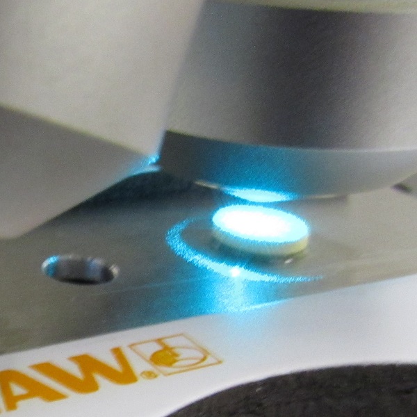
- How to reduce the effects of background fluorescence
- How the excitation wavelength affects the penetration depth into a sample
- The optimum power output to maximise signal levels while avoiding sample damage
- How focus type, optics, excitation wavelength and scanning affect laser spot size, sample coverage and spatial resolution
By the end of the webinar, you will understand how to select the optimum lasers for your samples.
Watch now
Part 2: What makes a good Raman spectrometer
- The optical layout of a dispersive Raman spectrometer
- How spectrometer design affects throughput and sensitivity
- How detector options affect optical efficiency
- Achieving both high spectral resolution and a broad spectral range
- Controlling the confocality of a Raman microscope
By the end of the webinar, you will understand the key factors that affect spectrometer performance, to help you choose the best system for your analyses.
Watch now
Part 3: Demonstrating how to use a Raman microscope
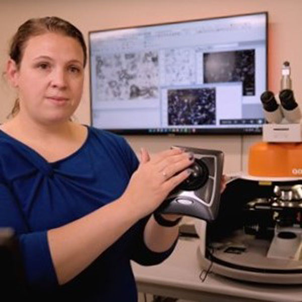
- Instrument layout and optics, from the excitation laser to the detector
- How the numerical aperture (N.A.) of microscope objectives affect sensitivity, spatial resolution and confocality
- Illumination options (brightfield, darkfield and polarised) for viewing different sample types
- Automated switching between lasers and the effect on Raman spectra
- Automated daily silicon calibration using an internal reference
- Sample holders and accessories for films, powders, gems, liquids and larger samples
- How Raman imaging can reveal spatial information
On completion, you will have learned not only how to collect good Raman data, but you will also have seen this demonstrated on a real instrument by our Raman experts.
Watch now
Further reading
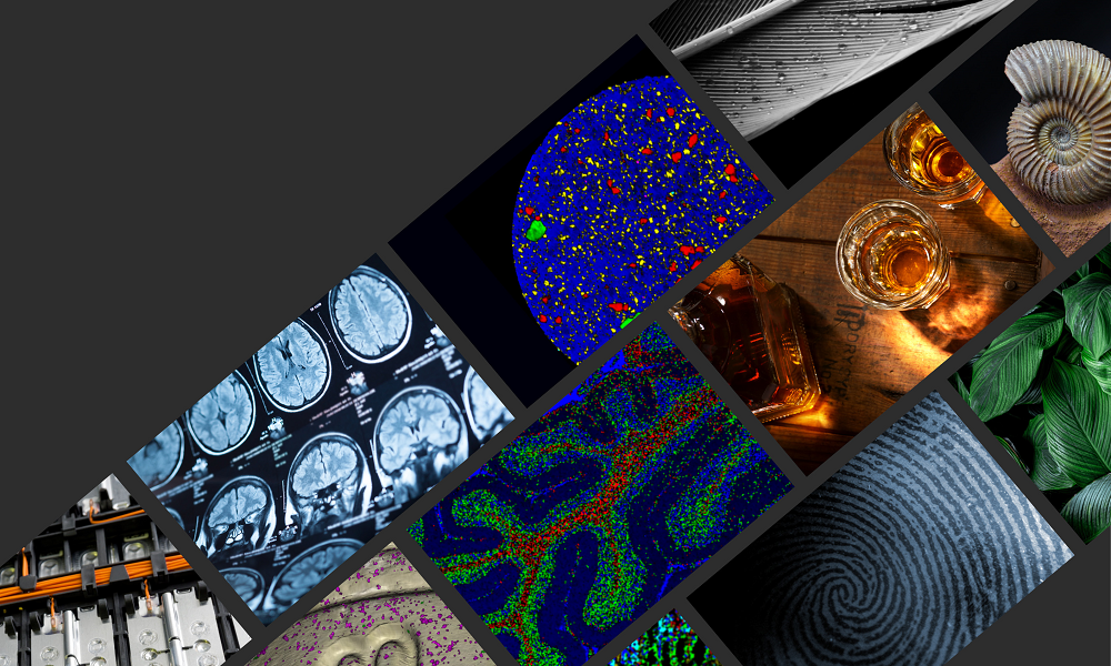
We have a whole range or articles, case studies and news stories about Raman spectroscopy.
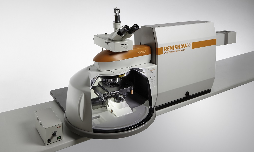
About the inVia microscope
Discover more about how the inVia confocal microscope is suitable for your organisations applications.