Study cell biology with Raman microscopy
Raman spectroscopy is a non-invasive and label-free technique. Extract chemical information without the need to manipulate genes, or use stains or antibodies. This helps ensure the results reflect the true chemistry of the cells.
Identify cells
Use Renishaw Raman systems to identify and distinguish, for example:
- cancer cells from normal cells
- stem cells from differentiated cells
- different sub-states in a cell population (e.g. stem and progenitor cells)
You can identify cells without known markers, based on their inherent chemical profiles. There is no need to conjugate with antibodies or manipulate genes.
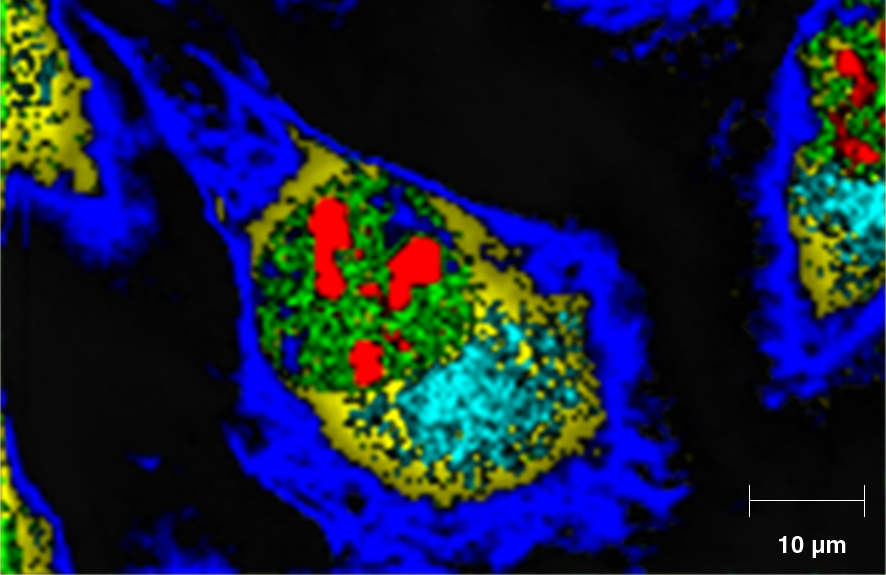
See fine biological detail
With high spatial resolution confocal Raman analyses you can examine:
- intracellular structures and biomolecules in individual cells, in situ
- the chemical contents of inclusions in yeast cells
- the lipid contents in cancer cells, to better understand lipid metabolism
Study individual cells within a population and determine cell-to-cell variability. For example, you can analyse the distribution of lipids and DNA in healthy and abnormal cells.
Study live cells
You can equip your Renishaw Raman system with a cell incubator. This chamber can control temperature, CO2 concentration, and humidity to keep cells in their normal physiological states during analysis.
Live cell data provides a better representation of any dynamic processes than end point experiments. For example, you can monitor the cells' response to changes in their environment or drugs. These responses may manifest themselves as metabolic or morphological changes, or cell death (apoptosis), all of which are detectable by Raman spectroscopy.
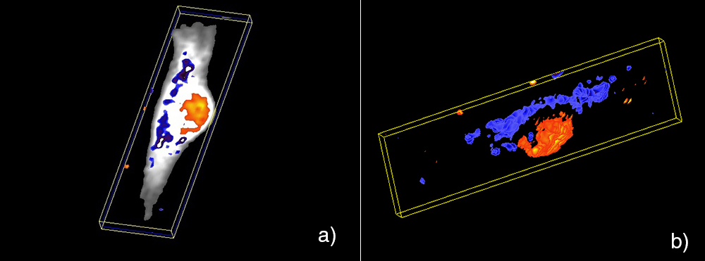
Resolve changes within cells in 3-D
Gather chemical information and produce 3-D views of your samples, and use these to verify the uptake of materials by cells. You can also determine the volumes of the cell and its organelles.
We're here when you need us
To find out more about this application area, or an application that isn't covered here, contact our applications team.
Contact our applications team
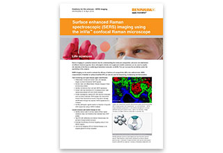
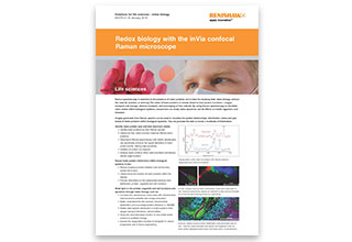
Webinar – Resonance Raman spectroscopy for redox biology research
Resonance Raman (RR) spectroscopy is the ideal tool for redox biology research. Not only does it demonstrate high sensitivity to haem proteins, it can also elucidate their oxidation and oxygenation states in situ (solution, organelles, cells and tissues). RR imaging provides both chemical and spatial information, enabling correlations between haem protein distribution, oxidation state, and protein/cell function to be made.
Watch the webinar
Download document


You may be interested in these papers:
Lau et al (2014) Biomedical Spectroscopy and Imaging 3: 237-247
McAughtrie et al (2013) Chem Sci 4: 3566-72
Kim et al (2010) Anal Bioanal Chem 398: 3051-3061