Correlative Raman microscopy with the Correlate™ software module
Combined Raman microscopy
The Correlate™ module enables the comparison of Raman results with many commonly used microscopy systems including Scanning Electron Microscopy (SEM), fluorescence, Atomic Force Microscopy (AFM), Infra-red and optical microscopes.
Features at a glance
- Coordinate Manager - To import and transform coordinates from commonly used microscopy systems to your Renishaw Raman system
- Image Alignment Tool - With translation, rotation, sizing and aspect ratio control for overlay with variable transparency
- Batch measurements - To automate the same Raman measurement at different positions on the sample
Easy to use
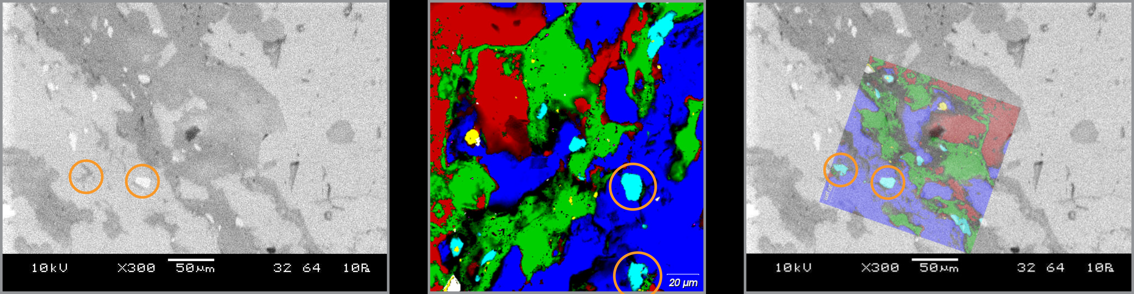
Simply record the coordinates of three or more reference points and data acquisition points on your sample. The Correlate module then guides you to the regions of interest on your samples after you have transferred them between microscopes. This enables you to get the data from the same locations and overlay the images for complementary interpretation.
Understand your sample with multimodal imaging
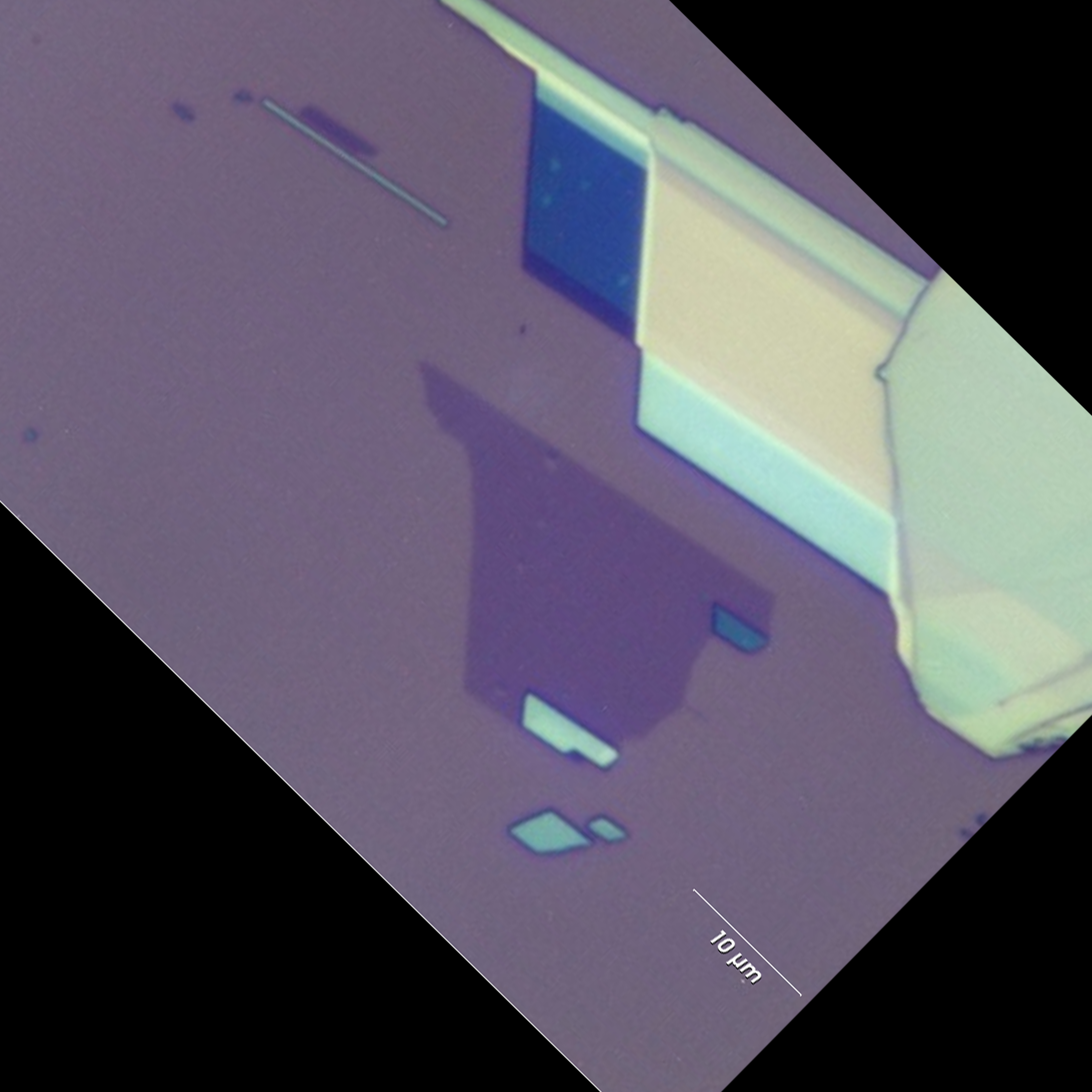
By combining the topographic details from the AFM image and images of different crystal domains from the white light image, you can gain a fuller understanding of the sample in one image.
The Correlate module is part of Renishaw's WiRE 5.3 software and can be used with the inVia confocal Raman microscope, the Virsa™ Raman Analyser, and the RA800 series.


Download document