Raman spectroscopy’s potential to identify diseased cartilage in osteoarthritis
Osteoarthritis is the leading cause of chronic disability in the elderly. In 2003, the World Health Organisation estimated that it affects 9.6% of men and 18% of women over the age of 60i. Patient treatment could be significantly improved if we could detect the first signs of cartilage degeneration earlier.
A group of researchers and clinicians at University College London, Royal Veterinary College and Charing Cross Hospital, led by Dr Riana Gaifulina, have investigated this. They have demonstrated that Raman spectroscopy could determine the grade of cartilage erosion and overall osteoarthritis disease state. This could lead to an earlier identification and treatment of osteoarthritic degeneration.
Rapid non-invasive Raman spectroscopy can detect small molecular changes in cartilage constituents. They looked for the target molecule, sulphated glycosaminoglycan (sGAG), in cartilage samples, using a Renishaw inVia™ confocal Raman microscope to acquire single spectra across the cartilage surface. This gave a comprehensive and representative analysis of the samples, so they could assess the progression of cartilage degradation. The researchers were able to identify the sGAG S-O stretching bond as an indicator of late stage cartilage damage by using Renishaw's WiRE™ curve fitting algorithm to analyse the spectral range, 980 cm-1 to 1120 cm-1 and, in particular, the 1063:1003 cm-1 peak ratio.
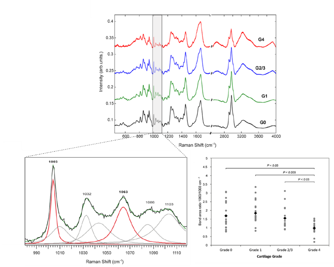
Raman spectra from Grade 0 to 4 cartilage samples. Curve fitting is applied over the 980 cm-1 to 1120 cm-1 spectral range. The 1063:1003 cm-1 peak ratio shows a significant difference for Grade 4 disease progression.
The researchers also performed principal component analysis (PCA) on both the high wavenumber and fingerprint spectral regions, and found that the high wavenumber region could more easily discriminate early disease.
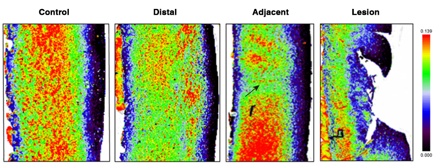
Then, knowing which peaks are indicative of disease, the researchers switched to the Renishaw RA816 Biological Analyser, as this is ideally suited to routine –use. They rapidly Raman imaged healthy and diseased areas of cartilage within multiple samples with Renishaw's fast StreamLine™ mapping technology. They found that Raman mapping could reveal significant differences between healthy cartilage and apparently normal looking cartilage from a lesioned joint, as well as the spatial distribution of cartilage constituents.
They also coupled a custom-built optical probe to the inVia system. This enabled them, for the first time, to explore the potential for in vivo in-clinic Raman analysis during knee joint arthroscopy. They acquired single point spectra from visually normal and lesioned cartilage within the same joint. Interpretable spectra could be acquired within 2 minutes. This work demonstrated that Raman data could be acquired in vivo from patients undergoing knee joint arthroscopy. However, it showed that analysis of single peak ratios is likely insufficient for predictive classification and more comprehensive multivariate spectral analysis is needed.
In this study the researchers showed that, due to its ability to detect small molecular changes in cartilage constituents, Raman spectroscopy may be an effective method to assess cartilage degeneration. This could allow clinicians to identify molecular alterations in osteoarthritic cartilage earlier which is necessary to more effectively treat this debilitating disease.
For further details on the paper in the Journal of Clinical Spectroscopy, please see: https://www.sciencedirect.com/science/article/pii/S2666054721000077
This is an open access article under the CC BY license (http://creativecommons.org/licenses/by/4.0/)
Disclaimer: The Renishaw inVia Raman microscope and RA816 Biological Analyser are designed for Research Use Only (RUO), not for use in diagnostic procedures.
Further reading
We have a whole range or articles, case studies and news stories about Raman spectroscopy.
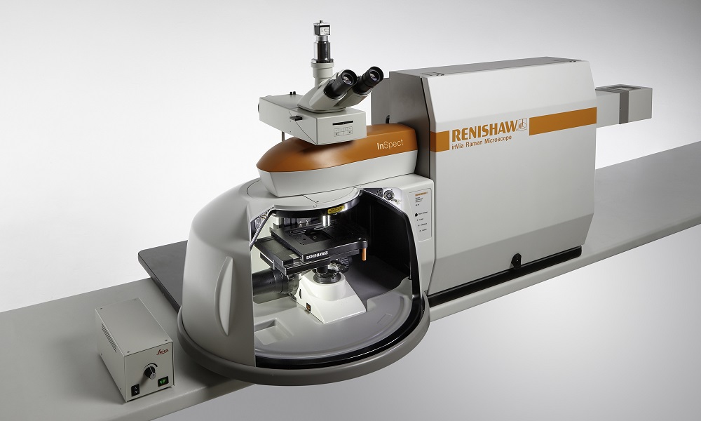
About the inVia microscope
Discover more about how the inVia confocal microscope is suitable for your organisations applications.