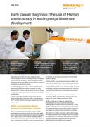
Early cancer diagnosis: The use of Raman spectroscopy in leading-edge biosensor development
Diagnosing most cancers at an early stage can have a profound impact on survival and life expectancy. If a malignant tumour has grown too big, or a cancer has spread, medical treatment becomes more difficult and the chances of surviving the disease are significantly reduced.
In a relatively short span of time, surface-enhanced Raman spectroscopy (SERS) has become a powerful tool in the development of innovative high-sensitivity biosensors capable of detecting and measuring cancer biomarkers at the earliest possible stage.
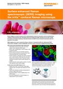
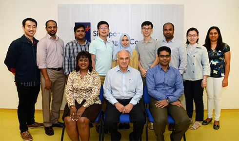
A pioneer in the field of SERS-based biosensor research, Singapore Bioimaging Consortium (SBIC) used Renishaw's inVia™ confocal Raman microscope to support this ground-breaking work.
SBIC's ground-breaking Raman spectroscopy bioimaging research
SBIC is a multidisciplinary biomedical research institute investigating human diseases, which are major public health issues, using molecular physiology and advanced bioimaging tools. It plays a pivotal role in strategic bioimaging projects and the development of biosensor technology. The institute is widely recognised for its ability to rapidly transfer results from its research into the clinical environment for the immediate benefit of patients.
Professor Patrick Cozzone, Executive Director of SBIC, said, “You could say that the work of SBIC relates directly to trade and industry. The rapid advancement of knowledge is an absolute pillar in what we do as a research institute, but so too is translating it into real commercial and economic outcomes - one that's inextricably linked to improving worldwide public health.”
One of the key measurement and analysis technologies employed in SBIC's ground-breaking research in the early detection of a range of different cancers is SERS.
SERS is a technique for molecular detection and characterization that relies on enhancing the Raman scattering signal of molecules that are adsorbed on, or near, SERS-active surfaces, such as nanostructured gold or silver.
The enhancement can be over a billion times in some cases and enables the analysis of low concentrations of material.
Since 2008, the institute's Laboratory of Bio-Optical Imaging has applied Renishaw's inVia confocal Raman microscope to the development of a wide range of pioneering SERS-based biosensor solutions that promise to achieve significantly cutting edge and more reliable cancer detection.
Biopolis, Singapore's R&D hub for biomedical science and home to SBIC
Develop biosensor platforms for early stage cancer diagnosis
Cancer represents one of the greatest public health challenges faced today. As reported by Cancer Research UK, in 2012, there were 14.1 million new cases worldwide and 8.2 million deaths from cancer. Early diagnosis of cancer is a vital factor for successful treatment. The research and development of biosensing solutions that enable reliable cancer diagnosis at the earliest possible stage is therefore an important international focus for national health system strategies and biomedical research institutes like SBIC.
Professor Malini Olivo, Head of the Laboratory of Bio-Optical Imaging at SBIC said, “The real challenge in detecting cancers at an early stage is that their pathological manifestations are simply not very evident. Probing a suspicious lesion by sight under even the best lighting can be highly unreliable, and sending a sample away to a laboratory for biopsy might take too long.”
She continued, “What's needed are highly sensitive, high-resolution cancer diagnostic techniques that can be used on the spot, enabling effective real-time management of early-stage cancers.”
Long proven as a highly accurate means of identifying, quantifying and differentiating between unknown materials in a wide range of industrial applications, Raman spectroscopy has in recent years been successfully applied to molecular diagnostic testing for the detection of cancer biomarkers.
The use of Raman spectroscopy in the investigation of biological tissues or fluids however presents a more demanding set of challenges. Biomarker expression levels in early stage cancers can be particularly low, making detection and monitoring more difficult. Using the SERS technique, the Raman signal can be amplified allowing for an increase in sensitivity.
A key focus of SBIC's research work has been to find innovative ways of improving and augmenting SERS-based solutions' performance and to develop biosensor platforms that are specifically optimised for early stage cancer diagnosis.
To achieve this, SBIC required a Raman microscope capable of operating reliably in a busy biomedical research laboratory environment.
Specifically, it needed to readily accommodate frequent change in research project teams, support high speed analyses for bulk screening programmes and deliver consistent high quality data output. Importantly, the microscope also needed to be durable and inherently stable in operation, regardless of the laboratory's uptime/downtime cycles.
How the Renishaw inVia Reflex Raman microscope is used
In its development of leading-edge SERS-based biosensor platforms for the early detection of cancers, SBIC used Renishaw's inVia confocal Raman microscope, a research grade microscope coupled to a high performance Raman spectrometer. The current configuration is an inVia Reflex with 633 nm and 785 nm laser, WiRE 4.3, chemometric software, StreamLine and StreamHR (high speed mapping).
Dr U.S. Dinish, Senior Research Scientist at SBIC, said, “A sensitive and specific spectrometer is important for supporting our wide and varied range of preclinical and clinical work, plus the development of SERS-based substrate, optical fibre and assay platforms. Other important considerations are ease-of-use, the availability of safety features, and the speed of data throughput.”
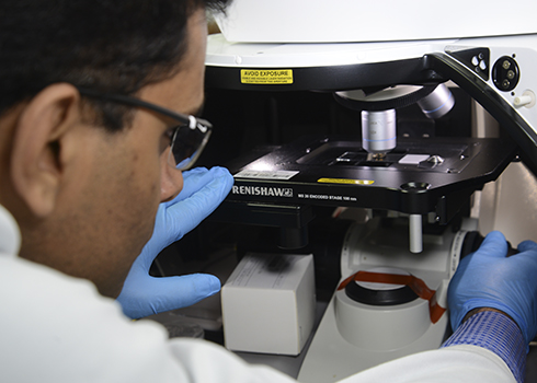 Raman spectrometer in action
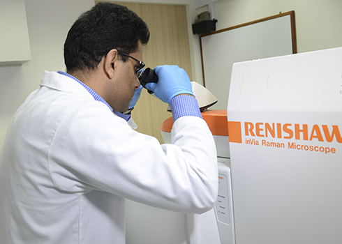
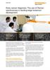
 Dr U.S. Dinish using the inVia confocal Raman microscope
A sensitive and specific spectrometer is important for supporting our wide and varied range of preclinical and clinical work, plus the development of SERS-based substrate, optical fibre and assay platforms. Other important considerations are ease-of-use, the availability of safety features, and the speed of data throughput.
Singapore Bioimaging Consortium (Singapore)
Development of photonic crystal fibre based SERS biosensor
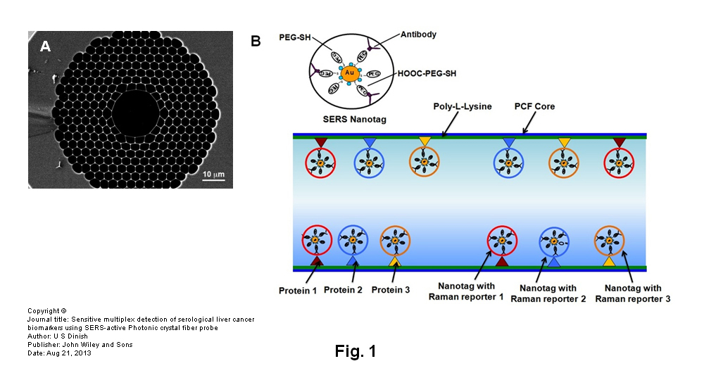
One of SBIC's key development projects that involves the use of the inVia Raman microscope and illustrates the scope of work undertaken in the labs combines SERS techniques with a hollow core photonic crystal fibre (HCPCF). Using this combination SBIC developed an ultrasensitive biosensor capable of multiplex detection of liver cancer biomarkers.
With its axially aligned air channels, the HCPCF provides an excellent platform for housing liquid and gaseous analytes for trace analysis. The HCPCF's light confinement characteristics and greater interaction length for analyte fluid and guided light created a biosensor with unprecedented sensitivity for samples as low in volume as 20 nL.
inVia's ease-of-use, flexibility and stability in delivering consistently reproducible spectral data was instrumental in achieving the biosensor's high detection sensitivity. Its option for fast switching between 633 nm and 785 nm lasers also ensured data throughput was maximised.
This joint development project between SBIC and XLIM, a joint research institute of the university of Limoges and CNRS (the French National Centre of Scientific Research), paves the way for the creation of next-generation cost-effective and portable biosensors for clinical and field-ready applications.
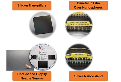
Examples of SERS-based substrate, optical fibre and assay
Ground-breaking biomedical performance
SERS-based biosensor research is providing completely new ways of detecting cancers at any early stage. The pioneering SERS techniques developed by the Laboratory of Bio-Optical Imaging at SBIC have the potential to increase the sensitivity and reliability of early stage cancer diagnosis, which could make a positive impact on patient survival rates.
In a diverse range of far-reaching SERS biosensor development projects, Renishaw's inVia confocal system demonstrated that commercially available research-grade Raman spectrometers offer the technical performance that is demanded by this vitally important area of biomedical research into human disease.
The inVia microscope has brought SBIC the user-friendliness and versatility, stability, sensitivity and fast spectral acquisition capability it needs in its work, empowering the institute to develop biosensor platforms in chips, assays and fibres in a variety of form factors. By helping to automate data acquisition processes in the laboratory's workflows, operational productivity has also been increased.
The above image is used under licence.
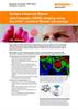
Figure 1 (A) SEM of the cross section of HCPCF.
Figure 1 (B) schematic of the binding of SERS nanotags to immobilized biomarkers inside the core of HCPCF for multiplex detection.
Copyright © (Journal title: Sensitive multiplex detection of serological liver cancer biomarkers using SERS-active Photonic crystal fiber probe / Author: U S Dinish / Publisher: John Wiley and Sons / Date: Aug 21, 2013)