Life sciences literature
Application notes
-
Application note: Tissue imaging with the inVia Raman microscope
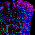
Raman tissue imaging is a unique method that can simultaneously describe the molecular composition and the distribution of multiple chemical species in tissues at a high spatial resolution, without labelling.
-
Application note: Redox biology with the inVia confocal Raman microscope
Raman spectroscopy is sensitive to the presence of haem proteins and is ideal for studying their redox biology, without the need for isolation or staining. The redox of haem proteins is closely linked to their protein functions – oxygen transport and storage, electron transport, and scavenging of free radicals. By using Raman spectroscopy to elucidate redox states within biological systems, researchers can study redox dynamics and its effects on health regulation and diseases.
-
Application note: Cell imaging with the inVia confocal Raman microscope
With Renishaw’s inVia confocal Raman microscope you can identify and characterise samples to provide chemical, spatial and structural information on multiple types of molecule, without labelling. It provides rich, detailed, chemical images and highly specific data at high spatial resolution, making it ideal for studying cells.
-
Application note: Surface enhanced Raman spectroscopy (SERS) imaging using the inVia confocal Raman microscope
Raman imaging is a powerful research tool for understanding the molecular composition, structure and distribution of different chemical species. Nano silver/gold colloids and roughened metallic substrates can be used to amplify the intensity of the Raman scattering of adsorbed molecules via SERS. This can increase the sensitivity and/or the specificity of the analysis. SERS imaging can be used to evaluate the efficacy of delivery of nanoparticles (NPs) into cells/animals. SERS measurement of labelled or surface-modified NPs can also be used for biosensing, multiplexing and theranostics.
Movie
-
Raman chemical imaging for life sciences
Short movie to demonstrate the benefits of using the inVia confocal Raman microscope for powerful, flexible, Raman Imaging for life sciences applications.
Webinar
You may also like to view our webinar - 'Resonance Raman spectroscopy for redox biology research'.
Application examples
We have also produced a range of application examples, including those below.
If you would like to find out more, please contact your local representative using the button below, quoting the relevant document reference.
Document reference | Description |
AS013 | 3D imaging a glioma cell. Reveal information about cells with inVia and 3D imaging. |
AS014 | 3D imaging rat spinal tissue. Reveal information about tissue with inVia and 3D imaging. In this example, the tissue components of rat spinal column are determined. |
AS016 | 3D imaging of cell organelles. Reveal information about cells with inVia and 3D imaging. In this example, the size and position of organelles wihin a glioma cell are determined. |
AS017 | Spermatozoon imaging. Reveal information about anatomical parts of organisms with inVia. In this example, the anatomical parts of a spermatozoon are determined. |
AS024 | AFM and 3D Raman imaging of glioma cells. Reveal detailed and complementary information on the composition of glioma cells by atomic force microscopy (AFM) and inVia 3D Raman imaging. |
AS025 | Fluoresence and 3D Raman imaging of cell organelles. Unravel detailed and complementary information on the composition of glioma cells using confocal 3D Raman and fluorescence imaging. |
AS031 | Distinguish embryonic stem/carcinoma cell lines without labelling. In this example, label free embryonic stem cell (ESC) and embryonic carcinoma cell (ECC) cell lines were differentiated using StreamLine™ imaging and principal component analysis. |
AS034 | Raman imaging of spermatozoa. Reveal anatomical and chemical in normal and abnormal cells with inVia. In this example, the anatomical parts of a phenotypically normal and a phenotypically abnormal spermatozoon are determined. |
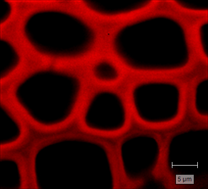
AS036 | Raman imaging of plant cell walls. Reveal the distribution and the relative thickness of plant cell walls and syringyl lignin using StreamLineHR imaging |
AS037 | Raman imaging of engineered melanoma models. Engineered 3D composites can be used to study the biochemistry of tumours, such as melanoma, and to investigate the impact of drugs on disease models. The tumour's size, chemistry, and its invasion and chemical alteration to the neighbouring tissue, are typically assessed. |
AS038 | Raman live cell imaging. Non-invasive live cell imaging |
AS053 | Raman imaging to reveal the morphological and biomolecular features in apoptotic cells. Reveal the morphology in apoptotic cells using confocal StreamLineHR imaging |