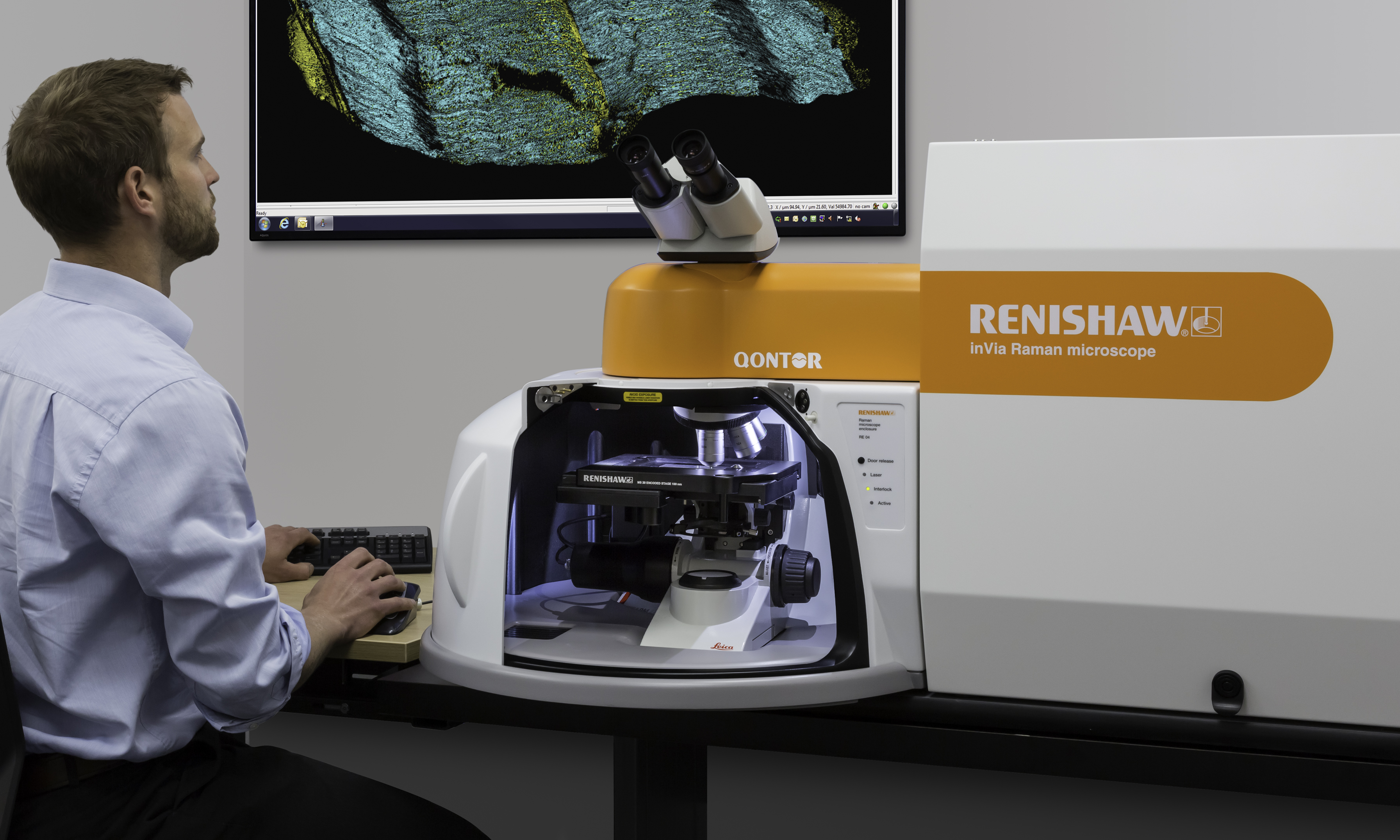
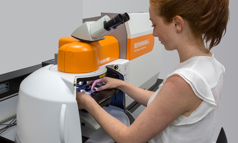
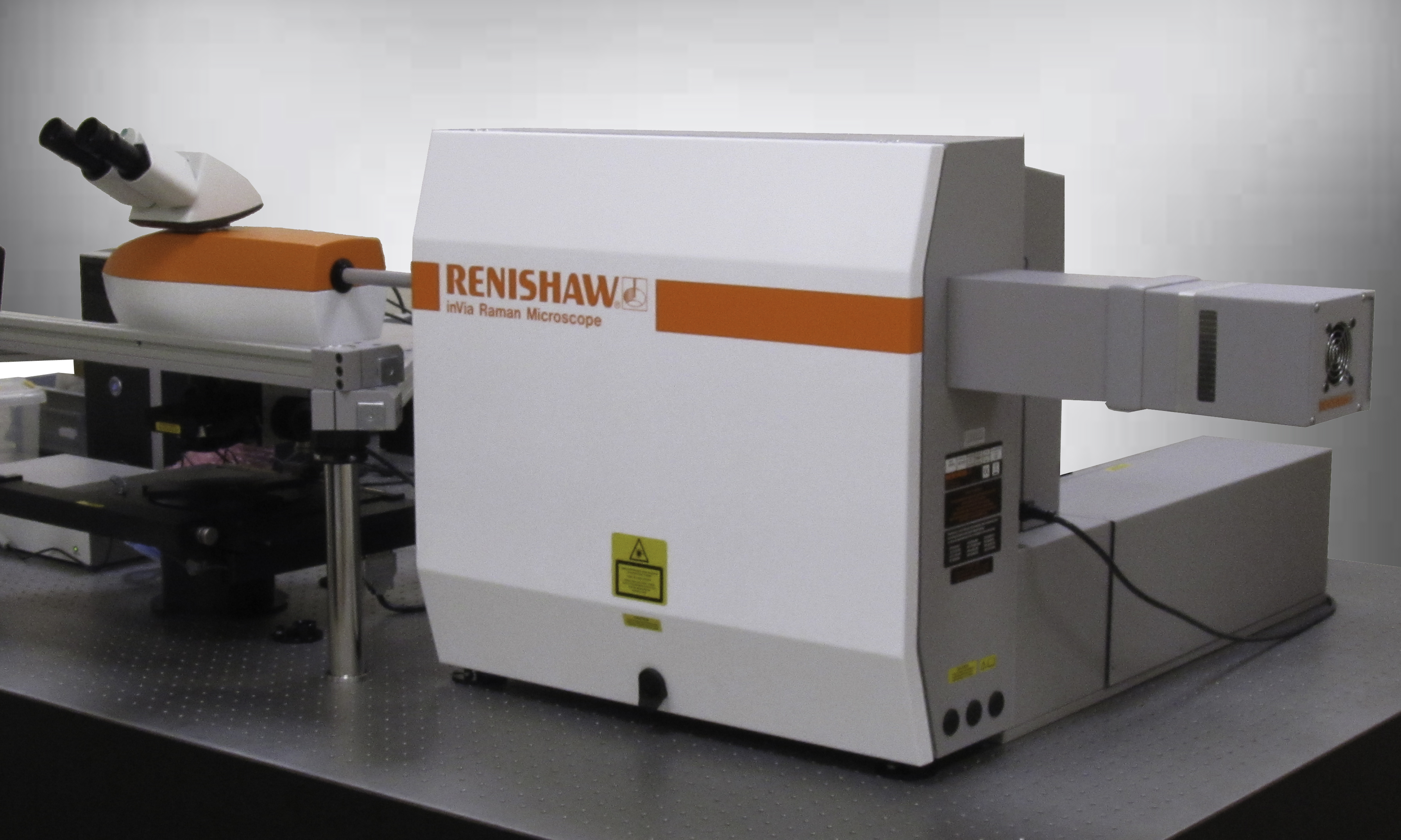
inVia™ confocal Raman microscope
The ultimate research-grade confocal Raman microscope delivers outstanding performance and the best data in the shortest time.
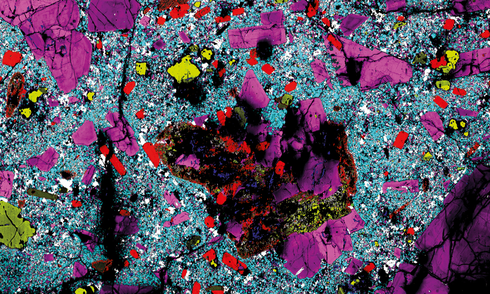
It is simple to operate yet delivers outstanding performance and reliable results, for even the most challenging experiments. You can produce both rich, detailed, chemical images and highly specific data from discrete points. With unparalleled flexibility, scientists and engineers, worldwide, trust the inVia microscope.
Features
The inVia microscope comprises a research-grade microscope coupled to a high-performance Raman spectrometer. It is simple to operate yet delivers outstanding performance—high signal throughput, combined with high spectral resolution and stability—giving reliable results, for even the most challenging measurements.
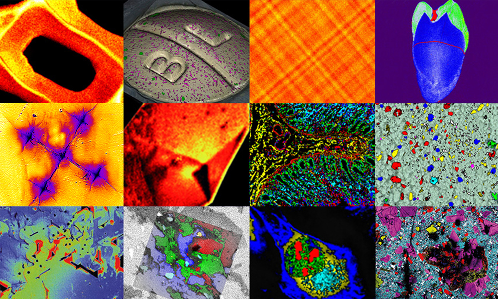
The inVia microscope's highly efficient optical design produces the best Raman data, even from minute traces of material. If you need to easily and reliably produce both rich, detailed, chemical images and highly specific data from discrete points, then the inVia microscope is the ideal system for you.
For more detailed information, download the inVia microscope brochure.
Speed and sensitivity
High sensitivity enables you to look at weak Raman signals and rapidly analyse even minute traces of material, monolayers and weak scatterers. It uses a stigmatic on-axis optical design which provides high optical efficiency, excellent stray light rejection and unparalleled sensitivity.
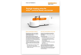
High spatial resolution
The optical design enables you to achieve a very high level of confocality (the ability to reject signal from regions away from the point of interest). This ensures you achieve high stability and the highest spatial resolutions possible, limited only by the inherent diffraction limit of the light.
High spectral performance
It can resolve spectral features narrower than 0.5 cm-1, so you can distinguish between close Raman bands, and differentiate very similar materials, such as pharmaceutical polymorphs. High stability allows you to monitor minute shifts in Raman band position (as low as 0.02 cm-1).
Exceptional stability
A custom designed honeycomb baseplate holds the inVia microscope and lasers in position and is so stable that an optical or anti-vibration table is not normally required. Key moving components are equipped with Renishaw high precision encoders which ensures everything is correctly placed.
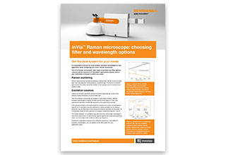
Spectral flexibility
You can configure the inVia microscope so that it is ideal for analysing your particular samples. It supports multiple lasers with automated computer-controlled switching. You can change excitation wavelength rapidly and quickly determine the best configurations to get reliable data from all your samples.
Sampling flexibility
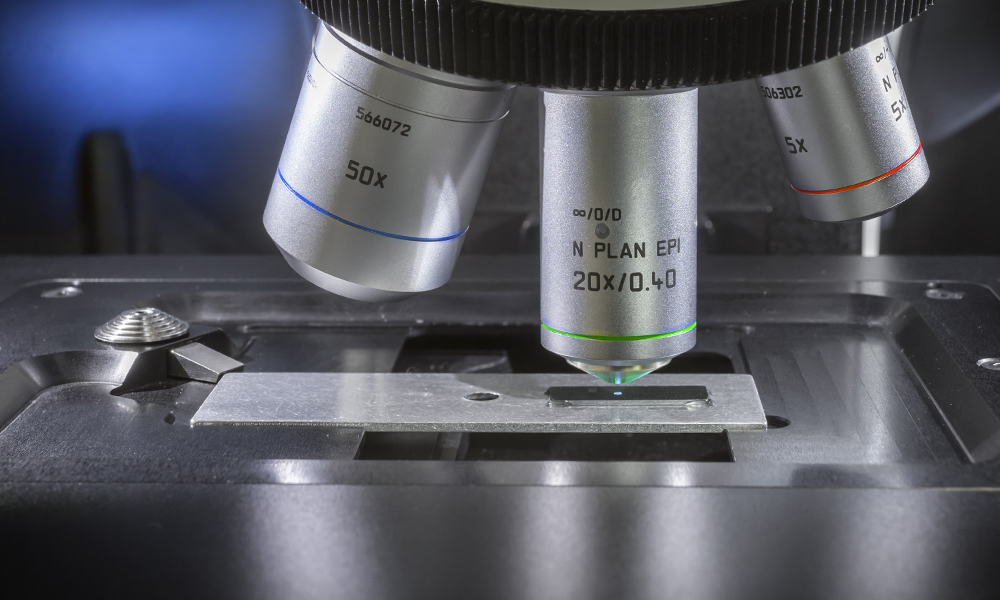
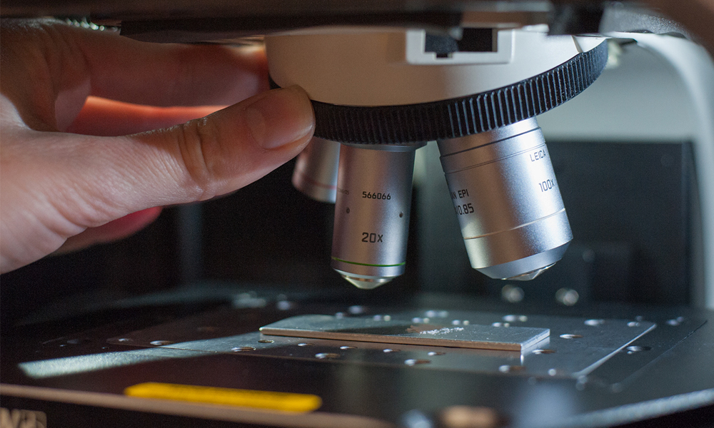
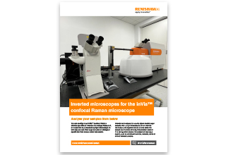
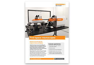
inVia supports both research-grade upright, inverted and open frame microscopes, as well as fibre optic probes for long distance remote analysis. It is compatible with a range of objective lenses and environmental cells enabling you to analyse your samples under a variety of environmental conditions.
Additional options

inVia™ InSpect
We have optimised our best-selling Raman microscope for use in forensic laboratories. The inVia InSpect microscope has the tools you need for non-destructive chemical analysis.
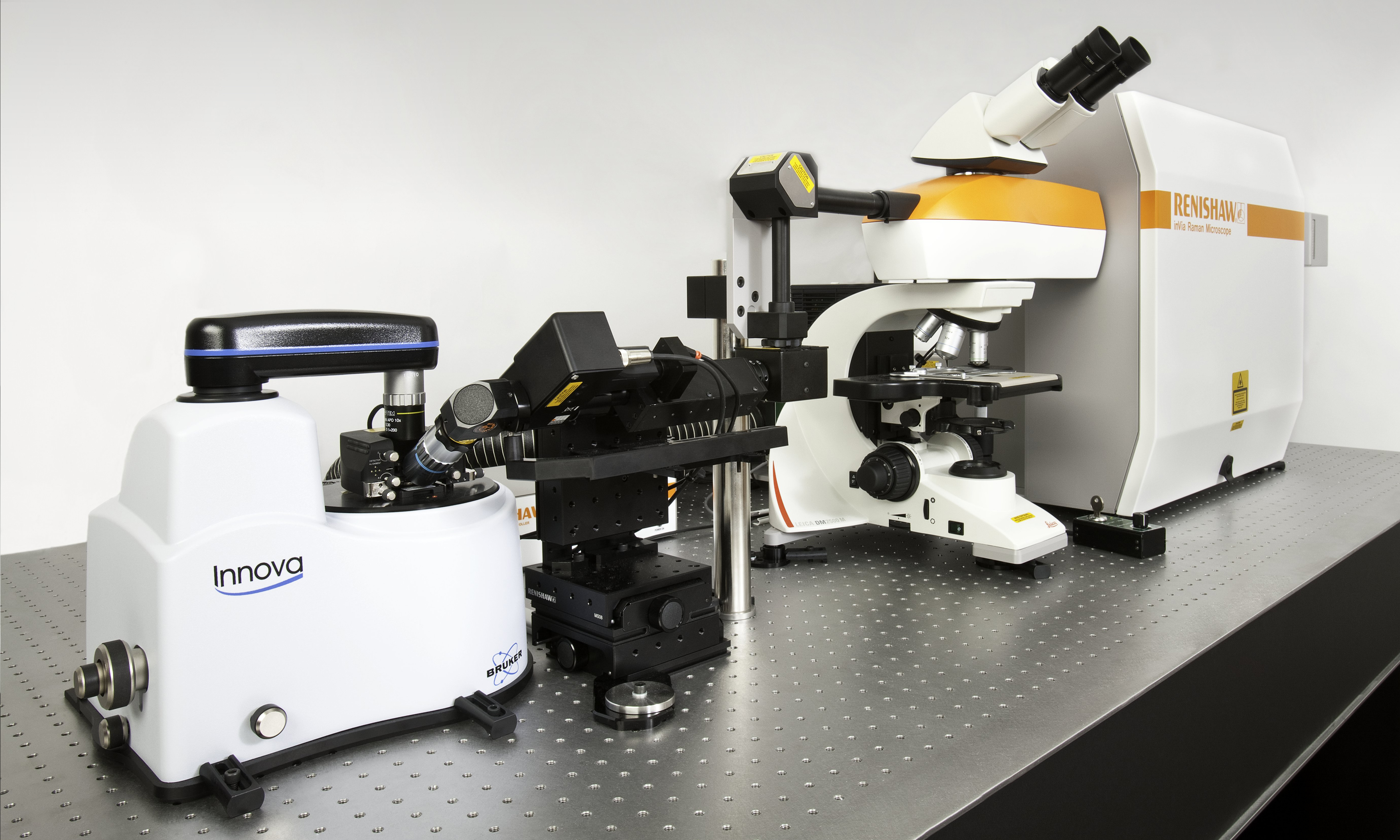
Combined Raman systems
Combine two or more analytical techniques in situ by coupling your Raman instrument with other complementary analytical instruments.
Custom Raman solutions
Our highly experienced Special Products Team can develop a custom solution to meet your specific requirements.
Applications
Applications for Raman spectroscopy
More and more laboratories are turning to Raman spectroscopy for chemical and structural characterisation. Raman analysis is non-destructive and non-contact, so it can be widely applied to almost any sample. Find out how you can apply the Raman technique to your application area.
System features and accessories
Our innovative Raman systems include many patented features to ensure that our Raman microscopes are fast, flexible and fully automated. Find out more about the system features, microscope components and accessories that help leading scientists to choose achieve great results.
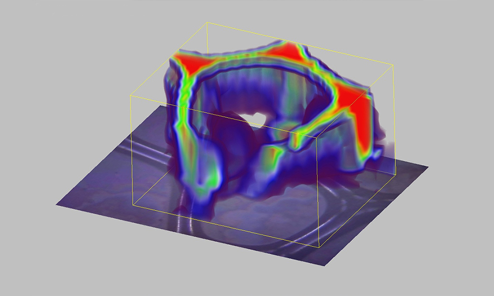
Maintain focus in real
time with LiveTrack
The inVia microscope uses LiveTrack™ automated focus tracking technology to acquire, in real-time, accurate and repeatable spectra and topography from samples with extensive variations in height. Create stunning 3D images of uneven, curved or rough surfaces without the need for pre-scanning. Watch the video to see an example.
LiveTrack technology
Analyse liquid or gas samples easily with the macro sampling kit
The macro sampling kit makes it easy to analyse liquid or gas samples in vials and cuvettes. Installed in seconds, it helps to get your Raman analysis done quickly.
Why choose a Renishaw Raman system
By choosing Renishaw products you can be confident you have made a sound investment. Your Raman system will be easy to use and will produce repeatable, reliable data, even from challenging samples. And it will last.
Want to find out more?
Your local representative will be happy to help with your enquiry. You can contact them by completing a form or sending an email.
Get our latest updates
Stay up-to-date with our latest news, webinars, application notes and product launches delivered directly to your inbox.
Download document

Specifications
The inVia microscope is available in three models; from the flagship inVia Qontor system - with full automation and focus tracking technology - to the entry level inVia Basis system.
Parameter | Value | |
| Wavelength range | 200 nm to 2200 nm | |
| Lasers supported | From 229 nm to 1064 nm | |
| Spectral resolution | 0.3 cm-1 (FWHM) | Highest typically necessary: 1 cm-1 |
| Stability | < ±0.01 cm-1 | Variation in the centre frequency of curve-fitted Si 520 cm-1 band, following repeat measurements. Achieved using a spectral resolution of 1 cm-1 or higher |
| Low wavenumber cut-off | 5 cm-1 | Lowest typically necessary: 100 cm-1 |
| High wavenumber cut-off | 30,000 cm-1 | Standard: 4,000 cm-1 |
| Spatial resolution (lateral) | 0.25 µm | Standard: 1 µm |
| Spatial resolution (axial) | < 1 µm | Standard: < 2 µm. Dependent on objective and laser |
| Detector size (standard) | 1024 pixel × 256 pixel | Other options available |
| Detector operating temperature | -70 °C | |
| Rayleigh filters supported | Unlimited | Up to four filter sets in automated mount. Unlimited additional filter sets supported by user-switchable accurately-locating kinematic mounts |
| Number of lasers supported | Unlimited | One as standard. Additional lasers beyond 4 require mounting on an optical table |
| Windows PC controlled | Latest specification Windows® PC | Includes PC workstation, monitor, keyboard and trackball |
| Supply voltage | 110 V AC to 240 V AC +10% -15% | |
| Supply frequency | 50 Hz or 60 Hz | |
| Typical power consumption (spectrometer) | 150 W | |
| Depth (dual-laser systems) | 930 mm | Dual laser baseplate |
| Depth (triple-laser systems) | 1116 mm | Triple laser baseplate |
| Depth (compact) | 610 mm | Up to three lasers (laser type dependant) |
| Typical mass (excluding lasers) | 90 kg |
Sample viewing | inVia Basis | inVia Reflex | inVia Qontor |
| Stereo viewing (binocular eyepieces) | ■ | ▲ | ▲ |
| Memorised and automatic post collection viewing | - | ▲ | ▲ |
| Software microscope control | - | ▲ | ▲ |
| Automatic white light/Raman switching | - | ▲ | ▲ |
| Automatic white light saving with data | - | ▲ | ▲ |
| Combined white light and laser video viewing | - | ▲ | ▲ |
| White light auto-focus (LiveTrack) | - | - | ▲ |
Raman data collection | inVia Basis | inVia Reflex | inVia Qontor |
| Automated measurement queuing | ▲ | ▲ | ▲ |
| Automatic focus tracking (LiveTrack) | - | - | ▲ |
Alignment and performance checking | inVia Basis | inVia Reflex | inVia Qontor |
| Internal neon wavelength calibration source | - | ▲ | ▲ |
| Internal reference standards for auto-calibration | - | ▲ | ▲ |
| Automated Raman calibration correction (quick calibration) | ▲ | ▲ | ▲ |
| Laser auto-align | ▲ | ▲ | ▲ |
| Raman signal auto-align | ▲ | ▲ | ▲ |
| Performance health check | - | ▲ | ▲ |
Key
| - not available | ■ option | ▲ included |