Multiple component 3D Raman volume image of a glioma cell. (jpg)
File size: 234 kB
Language: Language Independent
Dimensions: 1516 x 940 px
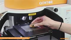
Latest videos - Raman spectroscopy
Didn't find what you were looking for?
Tell us what you couldn’t find and we will do our best to help.


