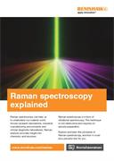
Raman image of human osteosarcoma (bone cancer) cells. (jpg)
File size: 289 kB
Language: Language Independent
Dimensions: 703 x 886 px
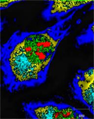
Raman image of human osteosarcoma (bone cancer) cells, showing the nuclei (green), nucleoli (red), membrane bound organelles (cyan) and the cell body (yellow – thick region, blue – membranous area). Renishaw thanks Dr Frederick Coffman, Rutgers New Jersey Medical School, USA, for providing the cell sample.
Latest videos - Raman spectroscopy



Latest items - Raman spectroscopy
Didn't find what you were looking for?
Tell us what you couldn’t find and we will do our best to help.